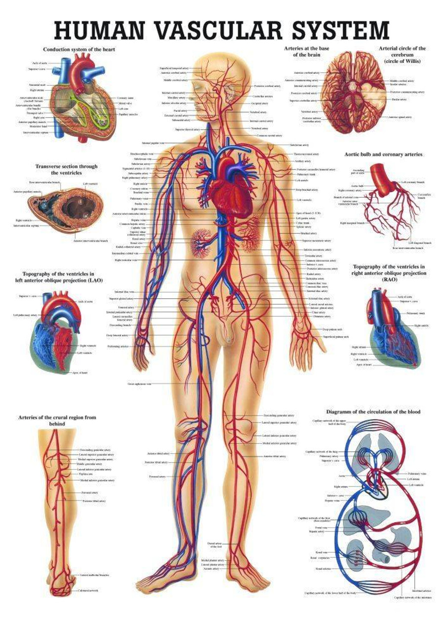
This is a detailed and educational anatomical chart focused entirely on the human liver. Printed on heavy paper, it measures 20 inches by 26 inches – a great size for clear visibility in a classroom, doctor's office, clinic, or even a student's study space. The main illustration shows an antero-visceral (front) view of the liver and its surrounding organs. Smaller, detailed diagrams break down various aspects: the liver's location in the chest cavity, the distribution of its vessels and ducts, the duct system highlighting common sites for gallstones, different liver segments from multiple views, the portal system, the structure of a liver lobule, and an illustration of cirrhosis. It's an excellent resource for medical students, nursing students, patients seeking to understand liver conditions, or educators in the health sciences. The version referenced here (ISBN 9781587791758) is laminated with grommets at the top corners, making it durable, easy to wipe clean, and ready to hang on a wall or chart rail. A must-have for clear visual reference in any educational or clinical setting dealing with human anatomy.