
No Prescription Needed: Best Over the Counter Orthotics for Flat Feet
July 28, 2026
Step Pain-Free Again: The Best Foot Tendonitis Relief Options
July 30, 2026
Relieving the Pressure with Thigh Fasciotomy
When Pressure Becomes an Emergency: Understanding Thigh Fasciotomy
A thigh fasciotomy is an emergency surgical procedure that cuts open the fascia — the tough tissue surrounding the thigh’s muscle compartments — to relieve dangerous pressure buildup before it causes permanent muscle and nerve damage.
Quick Answer: What Is a Thigh Fasciotomy?
| Question | Answer |
|---|---|
| What is it? | Emergency surgery to release pressure inside the thigh’s muscle compartments |
| Why is it needed? | Compartment syndrome cuts off blood supply to muscles and nerves |
| How urgent is it? | Critical — irreversible damage can occur within 6–8 hours |
| What happens without it? | Permanent muscle death, nerve loss, amputation, or death |
| What does it involve? | One or two surgical incisions along the thigh to open the fascia |
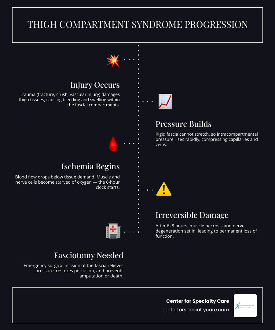
Every muscle in your thigh sits inside a tight fascial compartment. Normally, that’s fine. But after a severe injury — a crushing blow, a femur fracture, or a vascular trauma — fluid and blood can pour into those compartments faster than the rigid fascia can expand.
Pressure climbs. Blood flow drops. Within hours, muscle and nerve tissue begin to die.
This condition is called acute compartment syndrome, and it is a true surgical emergency. The stakes are high: studies show that over 50% of patients who develop thigh compartment syndrome experience long-term functional deficits, and mortality can reach as high as 47% without immediate intervention.
I’m Dr. Corey Welchlin, a board-certified orthopedic surgeon with over 30 years of experience treating complex musculoskeletal injuries at the Center for Specialty Care in Fairmont, Minnesota. Understanding when and how to perform a thigh fasciotomy is one of the most critical skills in orthopedic trauma surgery, and this guide walks through everything you need to know — from diagnosis to recovery.
Understanding Thigh Compartment Syndrome: Causes and Clinical Signs
To understand why a thigh fasciotomy is necessary, we must first look at the unique anatomy of the thigh and how easily things can go awry.
The human thigh is surrounded by a thick, unyielding muscular envelope. This envelope is divided into three distinct anatomical compartments: the anterior, posterior, and medial (adductor) compartments. Because the fascia enclosing these areas cannot easily stretch, any sudden increase in volume within a compartment elevates the internal pressure. If this pressure exceeds the capillary perfusion pressure, blood flow stalls, leading to rapid muscle ischemia (lack of oxygen) and tissue death.
What Triggers Acute Compartment Syndrome in the Thigh?
While the lower leg is the most common site for compartment syndrome (accounting for roughly 68% of civilian cases), the thigh is the site in approximately 9% of civilian cases requiring emergency decompression. In combat settings, thigh compartment syndrome represents about 8% of all fasciotomies.
Because the thigh is a large volume area and its compartments can occasionally bleed into the hip or buttocks, it takes a massive amount of energy or fluid accumulation to trigger compartment syndrome here. The most common causes we see include:
- Severe Femur Fractures: High-energy fractures can tear surrounding blood vessels, filling the compartment with blood.
- Crush Injuries: Heavy machinery accidents or motor vehicle collisions can directly crush muscle tissue.
- Vascular Trauma and Reperfusion Injury: A sudden restoration of blood flow after an arterial injury can cause severe swelling.
- Deep Vein Thrombosis (DVT): Severe blockages, such as an iliofemoral deep venous thrombosis, can cause massive venous congestion and drive fluid into the tissues.
- External Compression: Long periods of immobilization, tight casts, or restrictive wraps.
The risk of needing a fasciotomy varies widely by injury mechanism. For example, the incidence is just 0.9% after a standard motor vehicle collision but jumps to 8.6% after a gunshot wound. Furthermore, the rate ranges from 2.2% for patients with closed fractures to a staggering 41.8% for patients who suffer combined venous and arterial injuries.
If you or a loved one are recovering from a severe high-energy injury, our specialized team at the Center for Specialty Care provides expert Orthopedics evaluations to monitor and treat complex post-traumatic conditions.
Recognizing the Symptoms and the Fallacy of the Distal Pulse
Diagnosing compartment syndrome in the thigh relies heavily on clinical evaluation. Time is of the essence, and we look for the classic clinical indicators, often referred to as the “6 Ps”:
- Pain Out of Proportion: Pain that is far more severe than expected for the injury and does not respond to strong pain medications.
- Pain on Passive Stretch: Squeezing or stretching the affected muscles gently causes excruciating pain. For example, passive knee flexion will stretch the anterior compartment, provoking extreme distress.
- Paresthesia: Numbness, tingling, or a “pins and needles” sensation, indicating nerve irritation.
- Pallor: Pale, cold, or discolored skin.
- Paralysis: Inability to move the toes or flex the knee (a very late sign of severe damage).
- Pulselessness: The absence of a distal pulse.
The Distal Pulse Fallacy
It is a common and dangerous misconception that having a strong, palpable pulse in the foot means the thigh is safe. This is not true.
Because the main arteries running through the thigh are under high pressure, blood will continue to flow through them to the foot even when the pressure inside the muscle compartments is high enough to cut off the tiny capillaries supplying the muscles. By the time a patient actually loses their distal pulse, irreversible muscle death has already occurred. A pulse should never be used to rule out compartment syndrome.
Diagnostic Criteria and the Role of Pressure Monitoring
While a clear physical examination is the gold standard for diagnosing acute compartment syndrome, clinical signs can be difficult to evaluate in certain situations.
For instance, if a patient has a head injury, is under deep sedation, has a high spinal cord injury, or is pediatric, they cannot accurately report pain or sensory changes. In these challenging cases, we rely on objective intracompartmental pressure measurements.
When is Intracompartmental Pressure Monitoring Indicated?
Direct pressure monitoring is indicated when the patient is obtunded, unresponsive, or has a confounding neurological injury that makes a reliable physical exam impossible.
To measure this, we use a specialized device like a STIC catheter or a needle manometer inserted directly into the affected muscle compartment. To ensure accuracy, the needle must be placed within 5 cm of the fracture site or the area of maximal swelling.
For detailed clinical standards on managing these emergencies, you can review the Fasciotomy – StatPearls – NCBI Bookshelf.
Calculating Delta P for Thigh Fasciotomy Decision Making
To make the final decision for a surgical thigh fasciotomy, we calculate the “Delta P” ($\Delta P$), which is the difference between the patient’s diastolic blood pressure and the measured compartment pressure:
$$\Delta P = \text{Diastolic Blood Pressure} – \text{Compartment Pressure}$$
A $\Delta P$ of 30 mm Hg or less is highly indicative of compartment syndrome and serves as an immediate indication for emergency surgical decompression.
If a patient is hypotensive (has low blood pressure) due to trauma shock, their tissues are even more vulnerable to pressure. In these cases, we maintain a much higher index of suspicion, as irreversible damage can occur at even lower pressure thresholds.
| Compartment Status | Intracompartmental Pressure | Delta P ($\Delta P$) | Action Required |
|---|---|---|---|
| Normal | 0 to 10 mm Hg | N/A | Monitor clinically |
| Borderline / Suspected | 11 to 30 mm Hg | > 30 mm Hg | Close observation, serial exams |
| Acute Compartment Syndrome | > 30 mm Hg | $\le$ 30 mm Hg | Emergent Thigh Fasciotomy |
Surgical Techniques for Performing a Thigh Fasciotomy
Once the diagnosis of thigh compartment syndrome is confirmed, the patient must be taken to the operating room immediately. The primary goal of a thigh fasciotomy is complete surgical decompression of the affected muscle groups to restore vital blood flow.
Normally, a complete decompression of the thigh requires a lateral approach to release the anterior and posterior compartments. If the medial compartment remains tight, we perform a separate medial incision.
Step-by-Step Lateral Thigh Fasciotomy for Anterior and Posterior Decompression
The lateral approach is the workhorse of thigh decompression. It allows the surgeon to access both the anterior and posterior compartments through a single, long incision.
- Incision: We make a long longitudinal incision on the lateral side of the thigh, extending from the intertrochanteric line (near the hip) down to the lateral femoral epicondyle (near the knee).
- Expose the Fascia Lata: We dissect through the subcutaneous tissue to expose the glistening, tough fascia lata (iliotibial band).
- Anterior Decompression: We make a longitudinal incision along the length of the fascia lata, exposing the underlying vastus lateralis muscle of the anterior compartment.
- Posterior Decompression: To decompress the posterior compartment, we carefully retract the vastus lateralis muscle medially and superiorly. This exposes the lateral intermuscular septum. We then make a longitudinal incision through this septum to release the posterior compartment (hamstring muscles).
- Hemostasis: We carefully ligate or cauterize any perforating blood vessels to prevent deep hematomas from forming after closure.
For a deeper dive into the specific anatomical landmarks of this procedure, refer to Lower extremity fasciotomy techniques – UpToDate.
Medial Thigh Fasciotomy for Adductor Compartment Release
While the lateral incision decompresses the vast majority of cases, the medial (adductor) compartment must be evaluated independently. If the medial compartment remains tense and hard after the lateral release, a separate medial incision is mandatory.
- Incision: We place a longitudinal incision on the medial aspect of the thigh, starting approximately 5 cm distal to the groin crease and extending down toward the medial femoral epicondyle.
- Identify and Protect Key Structures: We must identify and carefully protect the great saphenous vein, the saphenous nerve, and the femoral artery and vein.
- Incision of the Fascia: We incise the fascia over the adductor muscles to completely relieve the pressure.
For an interesting clinical scenario involving multi-compartment lower limb decompression, you can read this URF Publishers Case Report.
Postoperative Wound Management and Rehabilitation
Performing the thigh fasciotomy is only the first step in saving the limb. Because the muscles swell significantly once the pressure is released, the surgical wounds cannot be closed immediately. They must be left open to heal in stages.
Temporary and Definitive Wound Closure Strategies
Directly after surgery, we apply temporary soft-tissue management. This prevents the skin edges from retracting too far while allowing the swelling to subside.
- Negative-Pressure Wound Therapy (NPWT / Wound VAC): We frequently apply a vacuum-assisted closure device. This uses continuous suction (usually set at 125 mm Hg) to pull excess fluid out of the wound, reduce swelling, and promote healthy granulation tissue.
- Dermatotraction (The “Shoelace” Technique): We can weave elastic vessel loops through skin staples along the wound edges. This maintains light tension on the skin, preventing it from shrinking while still allowing the muscle to swell safely.
- Delayed Primary Closure: Within 48 to 72 hours, we bring the patient back to the operating room for a “second look” to clean the wound and assess muscle viability. Once the swelling has gone down (usually within 5 to 7 days), we attempt to close the skin with sutures.
- Skin Grafting: If the swelling was severe and the skin edges cannot be brought back together safely, we apply a split-thickness skin graft (which is required in about 50% of all fasciotomy cases).
If you are undergoing or recovering from a major procedure, our surgical team at the Center for Specialty Care offers comprehensive Surgery and wound management options.
Preventing Volkmann’s Ischemic Contracture and Long-Term Complications
Leaving compartment syndrome untreated or delaying a fasciotomy past the critical 6-hour window can lead to devastating, permanent complications:
- Muscle Necrosis and Rhabdomyolysis: Dead muscle tissue releases toxins into the blood, which can cause life-threatening kidney failure.
- Volkmann’s Ischemic Contracture: When dead muscle tissue is replaced by stiff, non-elastic scar tissue, it permanently shortens the muscle, clawing the joints and causing severe deformity.
- Myositis Ossificans: Bone tissue abnormally grows inside the muscle, causing severe pain and stiffness.
- Infection: Dead muscle tissue is a perfect breeding ground for bacteria, often leading to deep bone infections or amputation.
To prevent these issues, our dedicated Post-Treatment Care programs focus on monitoring tissue healing and managing scarring.
Physical Therapy and Functional Recovery After Thigh Fasciotomy
Once the wounds are closed and healing, intensive physical therapy is crucial to restore mobility and strength. Over 50% of patients experience some long-term functional deficits, such as decreased knee flexion, sensory loss, or weakness.
Our customized Physical Therapy protocols are designed to combat these challenges through:
- Early Range of Motion (ROM): Gentle passive stretching to prevent joint stiffness and muscle contractures.
- Splinting: Keeping the knee in a semi-flexed position and the foot in a neutral position to prevent foot drop.
- Progressive Strengthening: Rebuilding the quadriceps and hamstrings once the wounds are fully healed.
- Scar Mobilization: Massaging the surgical scars to prevent them from sticking to the underlying muscle.
To help guide your recovery, here is a typical progression of rehabilitation exercises we use:
- Passive Range of Motion: Gentle, therapist-guided knee and hip movements to maintain joint flexibility.
- Isometrics: Contracting the quadriceps and hamstrings without moving the joint to prevent muscle wasting.
- Active-Assisted ROM: The patient begins moving the leg with light assistance.
- Progressive Resistance Training: Using light bands and weights to rebuild functional strength.
Frequently Asked Questions about Thigh Decompression
What is the recommended timing for a thigh fasciotomy?
A thigh fasciotomy should be performed as soon as the clinical diagnosis of compartment syndrome is made. The golden window to prevent permanent damage is within 6 hours of the onset of ischemia.
Waiting longer than 6 to 12 hours drops the rate of normal functional recovery to just 8% and significantly increases the risk of amputation and death. For more information on emergency timing, please see the Fasciotomy Protocols.
What are the anatomical compartments of the thigh?
The thigh is divided into three major compartments:
- Anterior Compartment: Contains the quadriceps femoris, sartorius muscle, and femoral nerve. It is responsible for knee extension and hip flexion.
- Posterior Compartment: Contains the hamstring muscles (biceps femoris, semitendinosus, semimembranosus) and the sciatic nerve. It is responsible for knee flexion and hip extension.
- Medial Compartment: Contains the adductor muscles (gracilis, adductor longus, brevis, and magnus) and the obturator nerve. It is responsible for pulling the leg inward.
To compare these to the compartments of the lower leg, you can read about Single and Dual-Incision Fasciotomy of the Lower Leg.
What are the long-term functional outcomes after a thigh fasciotomy?
While a fasciotomy is a lifesaving and limb-saving procedure, the initial trauma and the surgery itself can leave lasting impacts. More than half of patients experience long-term symptoms, including:
- Decreased knee flexion range of motion.
- Mild muscle weakness in the quadriceps or hamstrings.
- Numbness or altered sensation near the surgical scars.
- Chronic pain.
If you are dealing with persistent post-surgical pain, our Pain Management specialists are here to design a personalized plan to improve your quality of life.
Conclusion
A thigh fasciotomy is a highly technical, high-stakes procedure where every minute counts. Recognizing the early signs of compartment syndrome — and understanding that a normal pulse does not mean a limb is safe — is vital to preventing permanent disability.
At the Center for Specialty Care, we are committed to providing world-class, personalized orthopedic care to our communities in Fairmont, MN, Estherville, IA, Buffalo Center, IA, and St. James, MN. Whether you are recovering from a complex trauma, need expert physical therapy, or require specialized pain management, our team is here to support you every step of the way.
If you or a loved one are recovering from a traumatic lower limb injury or experiencing persistent leg pain, contact our Fairmont clinic today to schedule a comprehensive evaluation with our orthopedic specialists. Learn more about how we handle these critical situations by reading about Pillar/Fasciotomy: A Lifesaving Procedure for Treating Compartment Syndrome.