The Ultimate Guide to Custom Orthotics for Flat Feet
August 4, 2026
How to Do Exercises for Tendonitis in Foot Without Tripping Up
August 6, 2026
In Depth Guide to Fasciotomy Procedure and Recovery
When Every Minute Counts: What a Fasciotomy Is and Why It Matters
A fasciotomy is an emergency surgical procedure that cuts open the fascia — the tough tissue surrounding your muscles — to relieve dangerous pressure buildup inside a muscle compartment. Without it, that pressure can cut off blood flow and permanently destroy muscle and nerve tissue.
Quick answer: What is a fasciotomy?
- What it is: A surgery that opens the fascia to release pressure inside a muscle compartment
- Why it’s done: To treat acute compartment syndrome before muscles and nerves are permanently damaged
- When it’s needed: Most often after fractures, crush injuries, severe burns, or high-energy trauma
- Most common sites: Lower leg and volar forearm
- Time-sensitive: Must ideally be performed within 6 hours of symptom onset for the best chance of full recovery
This is not an elective procedure. It is one of the most urgent operations in orthopedic and trauma surgery. Delay can mean the difference between full recovery and permanent disability — or worse.
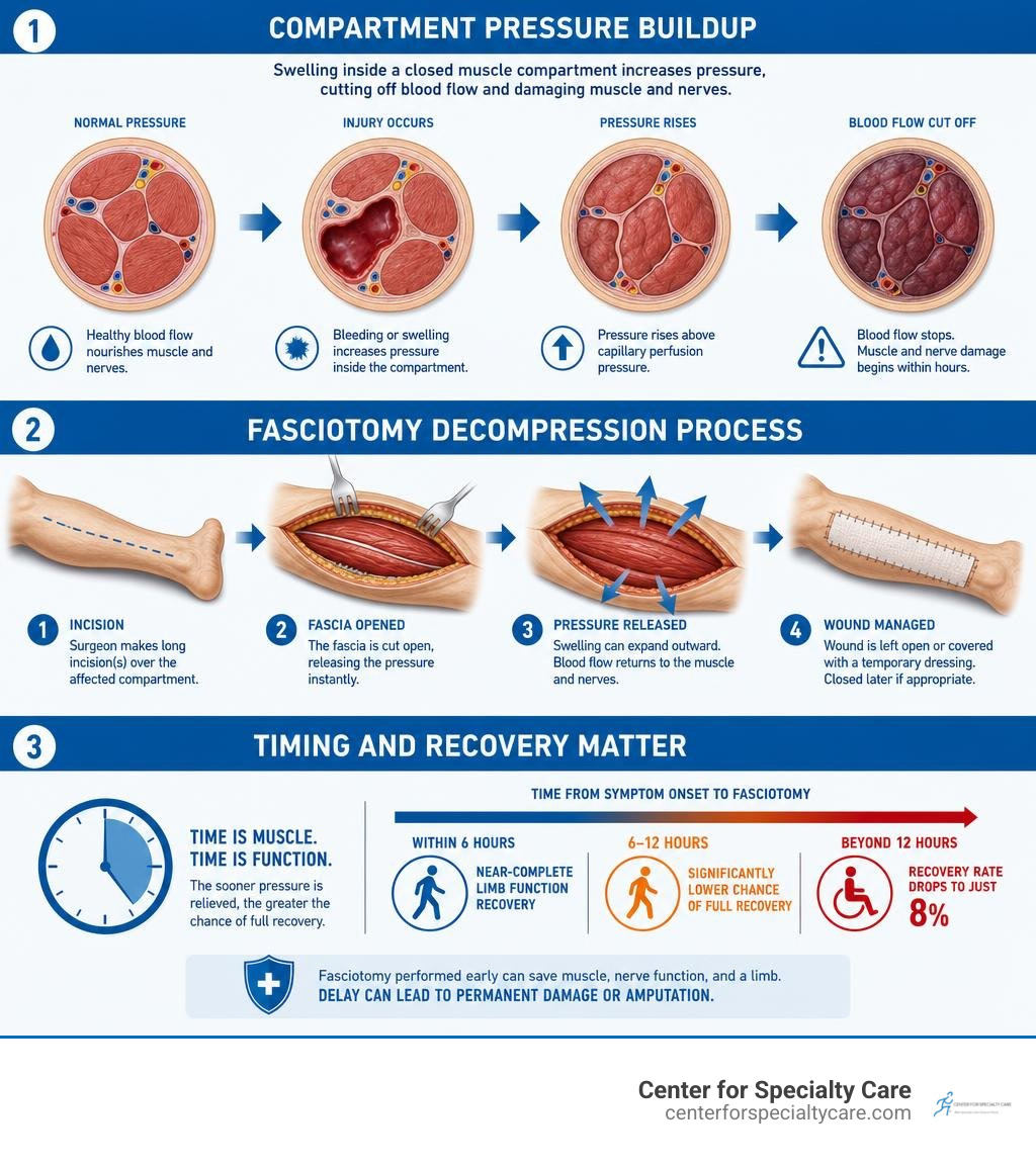
The numbers make that clear. Fasciotomies performed within 6 hours lead to near-complete limb function recovery. Wait beyond 12 hours, and that recovery rate drops to just 8%.
I’m Dr. Corey Welchlin, a board-certified orthopedic surgeon with over 30 years of experience treating acute and complex musculoskeletal conditions — including compartment syndrome requiring fasciotomy — at the Center for Specialty Care in Fairmont, Minnesota. In this guide, I’ll walk you through everything you need to know about this critical procedure, from how it works to what recovery looks like.
Understanding Compartment Syndrome and the Need for a Fasciotomy
To understand why a fasciotomy is necessary, we first have to look at human anatomy. Muscle groups in our arms and legs are organized into distinct anatomical units called compartments. Each compartment is encased by a dense, non-compliant layer of fibrous connective tissue known as fascia. While fascia provides structure and helps anchor muscles, it has almost no elasticity. It cannot stretch.
When an injury occurs within a closed compartment, bleeding or tissue swelling causes the internal pressure—known as intracompartmental pressure—to surge. As this pressure climbs, it eventually exceeds tissue perfusion pressure, which is the pressure required to keep blood flowing through tiny capillary networks. When intracompartmental pressure eclipses capillary perfusion, capillary collapse occurs. Blood supply shuts off, preventing oxygenated blood from reaching muscle tissue and peripheral nerves. The result is rapid, progressive muscle ischemia and cell death.
To identify this dangerous spike in pressure, clinicians closely monitor key clinical signs often referred to as the “6 Ps”:
- Pain out of proportion: Severe, deep, agonizing pain that is entirely disproportionate to the physical appearance of the injury and unmanaged by standard pain medications.
- Pain on passive stretch: Exquisite pain triggered when the clinician gently stretches muscles within the affected compartment.
- Paresthesia: “Pins and needles” sensation or numbness signaling early nerve dysfunction.
- Pallor: Pale skin, decreased temperature, or abnormal capillary refill in the extremity.
- Paralysis: Inability to move fingers or toes, indicating severe, advanced ischemia.
- Pulselessness: Absence of distal pulses, which is actually a late and grave sign (capillaries collapse long before major arteries lose pulsation).
When physical exams are inconclusive—such as when a patient is unconscious, sedated, or unresponsive due to severe trauma—we measure intracompartmental pressure directly using a needle pressure monitor. Diagnosis relies heavily on the “delta pressure” threshold: calculating the difference between the patient’s diastolic blood pressure and the compartment pressure. If the absolute compartment pressure is greater than 30 mmHg, or if the delta pressure drops to 20 mmHg or less, immediate surgical intervention is mandated.
As highlighted in the medical reference Fasciotomy – StatPearls – NCBI Bookshelf, surgical decompression remains the gold-standard treatment. At the Center for Specialty Care, we consider fasciotomy a lifesaving procedure for treating compartment syndrome because prompt pressure relief halts the cascade of muscle necrosis and permanent nerve destruction.
Anatomical Locations and Clinical Indications
While compartment syndrome can theoretically manifest in any enclosed anatomical space, it occurs most frequently in specific extremity compartments. By far, the lower leg is the most common site, followed by the forearm. However, we also treat compartment syndrome in the thigh, foot, hand, and buttock.

High-energy trauma—such as motor vehicle collisions, industrial accidents, and high-impact sports injuries—is the leading trigger for acute compartment syndrome. Closed long-bone fractures (particularly tibial shaft fractures) account for a substantial percentage of all cases. Other non-fracture triggers include severe crush injuries, high-voltage electrical burns, circumferential thermal burns, snakebites, tight cast applications, prolonged positioning during prolonged surgeries, and reperfusion following vascular repair.
In addition to acute trauma, athletes and military personnel occasionally experience chronic exertional compartment syndrome (CECS). Unlike acute trauma, CECS stems from exercise-induced muscle swelling within tight fascial borders, leading to activity-related pain, tightness, and cramping that resolves with rest. While initial CECS management involves conservative care, refractory cases may require surgical decompression, such as relieving the pressure with thigh fasciotomy or leg fascial release.
Timing Constraints and Relative Contraindications
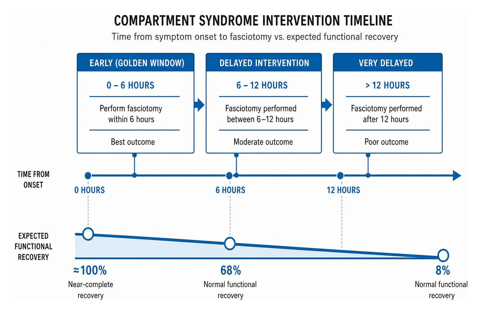
When acute compartment syndrome strikes, time is the ultimate variable. Nerve tissue begins suffering irreversible functional impairment within 2 to 4 hours of complete ischemia, and irreversible muscle necrosis begins at approximately 6 hours. Therefore, performing a fasciotomy within the early “golden window” (under 6 hours from symptom onset) yields an exceptional outcome, with near-complete functional limb recovery.
When surgical decompression occurs between 6 and 12 hours after onset, normal functional recovery drops to approximately 68%. If intervention is delayed beyond 12 hours, the prognosis becomes grim: only 8% of patients regain normal limb function.
This steep drop-off introduces major clinical and ethical questions regarding delayed presentation. Performing a fasciotomy on a limb that has suffered complete ischemic death for more than 12 to 24 hours presents serious relative contraindications:
- Reperfusion Injury: Opening blood flow to dead, infarcted muscle releases massive quantities of potassium, myoglobin, and inflammatory cytokines into the systemic circulation, triggering lethal cardiac arrhythmias and acute renal failure.
- Systemic Infection and Sepsis: Incising non-viable, necrotic muscle tissue exposes dead tissue directly to environmental pathogens, dramatically increasing the risk of overwhelming wound infection, sepsis, and amputation.
In battlefield trauma studies, delayed fasciotomies (>12 hours) resulted in twice the rate of amputation and triple the mortality rate compared to early interventions. In situations with late presentation and non-viable tissue, non-operative management or primary amputation may be recommended over delayed fascial release to safeguard the patient’s life.
Surgical Techniques and Procedural Steps
Executing a successful fasciotomy requires rapid preparation, precise anatomical knowledge, and meticulous operative execution. When acute compartment syndrome is confirmed, our surgical team immediately mobilizes the operating room.
Prior to incision, the team performs the standard World Health Organization (WHO) Surgical Safety Checklist. The patient is positioned supine, and regional or general anesthesia is administered. The entire affected extremity is scrubbed, prepped with antiseptic solution, and draped widely past the joint lines to allow full access. Proper equipment—including major orthopedic surgical trays, electrocautery, self-retaining retractors, skin markers, and vascular clips—must be fully prepared.
At the Center for Specialty Care, our board-certified surgeons work seamlessly alongside specialized surgical nurses and anesthesiologists to provide urgent surgery with precision and speed, following evidence-based protocols outlined in sources like Lower extremity fasciotomy techniques – UpToDate.
Leg Fasciotomy: Single-Incision vs. Double-Incision Approaches
The lower leg contains four distinct non-communicating compartments:
- Anterior Compartment: Contains tibialis anterior, extensor hallucis longus, extensor digitorum longus, and the deep peroneal nerve.
- Lateral Compartment: Contains peroneus longus, peroneus brevis, and the superficial peroneal nerve.
- Superficial Posterior Compartment: Contains gastrocnemius, soleus, and plantaris muscles.
- Deep Posterior Compartment: Contains tibialis posterior, flexor hallucis longus, flexor digitorum longus, and the posterior tibial neurovascular bundle.
To successfully decompress the lower leg, all four compartments must be released. Surgeons choose between two primary methods: the single-incision technique and the double-incision technique.
| Feature / Technique | Single-Incision (Davey/Rorabeck) | Double-Incision (Mubarak/Harges) |
|---|---|---|
| Incision Location | Single lateral incision along the fibular shaft axis | Two parallel incisions: anterolateral and posteromedial |
| Compartment Decompression | Anterior, lateral, superficial & deep posterior via one opening | Anterior/lateral via outer incision; superficial/deep posterior via inner incision |
| Technical Difficulty | Anatomically challenging; deep posterior release can be difficult | Standard approach; widely preferred due to clear anatomical exposure |
| Nerve Risk | Superficial peroneal nerve injury during deep dissection | Superficial peroneal nerve laterally, saphenous vein/nerve medially |
| Wound Tension | Concentration of wound tension along a single long incision | Pressure and tension distributed across two separate surgical sites |
The double-incision approach (Mubarak technique) is the most widely performed approach due to its safety margin and direct exposure:
- Anterolateral Incision: Placed halfway between the fibular shaft and anterior tibial crest. The surgeon carefully identifies the intermuscular septum separating the anterior and lateral compartments, making longitudinal incisions in both directions while protecting the superficial peroneal nerve.
- Posteromedial Incision: Placed 2 cm posterior to the posterior border of the tibia. Skin and subcutaneous tissues are incised, taking care to protect the saphenous nerve and great saphenous vein. The superficial posterior fascia is released longitudinally, followed by detaching the soleal bridge to expose and split the fascia of the deep posterior compartment.
Forearm Fasciotomy: Volar and Dorsal Techniques
Although upper extremity cases account for roughly 20 percent of all extremity fasciotomies, forearm compartment syndrome demands equal urgency to preserve fine motor function and hand dexterity. Approximately 1 percent of overall extremity trauma patients require an upper extremity fasciotomy. The forearm houses four primary functional spaces: superficial volar, deep volar, dorsal, and the mobile wad.
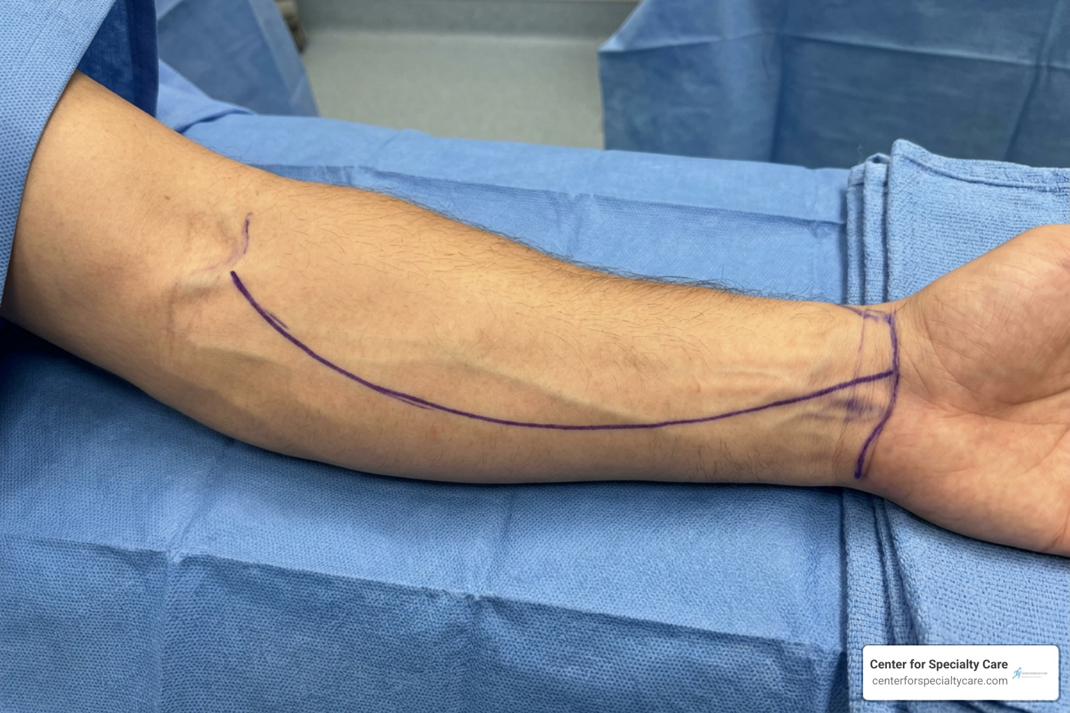
According to specialized clinical protocols detailed in Upper extremity fasciotomy techniques – UpToDate, decompressing the upper extremity requires step-by-step anatomical precision:
- Volar Incision: A curvilinear, extensile incision begins proximal to the antecubital fossa, passes diagonally across the flexor muscle belly, curves distally toward the ulnar border of the mid-forearm, and curves back across the wrist toward the palmar crease.
- Carpal Tunnel Release: The incision extends across the wrist flexor crease to release the transverse carpal ligament, preventing secondary median nerve compression within the carpal tunnel.
- Fascial Release: The superficial volar fascia is split along its entire length. Surgeons then retract the flexor carpi radialis and median nerve to directly open the deep volar compartment fascia.
- Dorsal Incision: If dorsal compartment pressures remain elevated, a straight longitudinal incision is made along the posterior forearm between the extensor digitorum communis and extensor carpi radialis brevis, releasing the dorsal compartment fascia and mobile wad.
Throughout both leg and arm techniques, soft tissue handling must be incredibly gentle to protect cutaneous nerves, including the superficial peroneal, saphenous, radial, and ulnar nerves.
Postoperative Management and Wound Closure Options
The end of the initial surgical procedure is not the end of treatment. Following a fasciotomy, muscle tissue surges through the opened incision as built-up tissue pressure drops. Consequently, the surgical wounds cannot—and should not—be closed primarily during the first operation. Attempting to force the skin closed would simply recreate the elevated compartment pressure.
Instead, the surgical site is dressed, supported, and reassessed within 48 to 72 hours. Our surgical team monitors tissue viability, resolves lingering swelling, and chooses an appropriate wound management strategy as part of comprehensive post-treatment care:
- Delayed Primary Closure: If muscle swelling subsides rapidly over 3 to 5 days, the wound edges can sometimes be pulled together and sutured cleanly without excess tension.
- Vessel Loop “Shoelace” Technique: Elastic vessel loops are threaded back and forth across the open wound through skin staples, mimicking a shoelace. Everyday adjustments gradually draw the skin margins together as swelling decreases, minimizing the need for skin grafting.
- Negative Pressure Wound Therapy (NPWT): Also known as vacuum-assisted closure (VAC), NPWT uses specialized foam dressings connected to gentle continuous suction. NPWT reduces local edema, stimulates granulation tissue formation, protects against external bacterial contamination, and pulls wound edges together.
- Split-Thickness Skin Grafting (STSG): In approximately 50% of fasciotomy wounds, massive tissue expansion prevents full primary skin closure. Once granulation tissue forms, thin layers of healthy skin are harvested from a donor site (typically the thigh) and grafted across the remaining surgical defect.
Potential Complications and Systemic Risks
A fasciotomy is an extensive, aggressive surgical procedure executed under high-stress emergency conditions. While it rescues limbs, patients and clinicians must remain vigilant regarding potential complications:
- Rhabdomyolysis and Acute Kidney Injury: As ischemic muscle tissue recovers or breaks down, damaged muscle cells release myoglobin into the blood stream. High levels of circulating myoglobin clog kidney tubules, leading to severe renal failure requiring intensive fluid hydration or hemodialysis.
- Infection and Sepsis: Leaving surgical incisions widely open creates a portal for microbial contamination. Deep tissue infections or osteomyelitis demand antibiotic therapy and repeat surgical debridement.
- Nerve and Vascular Injuries: Emergency dissections in swollen, distorted tissues carry risk of accidental nerve injury, leading to permanent sensory numbness or motor weakness (such as foot drop from superficial or deep peroneal nerve damage).
- Joint Stiffness and Scar Tethering: Extensive scarring across joint flexor lines can bind tendons and restrict range of motion, causing permanent joint contractures.
- Need for Revision Surgery: Incomplete decompression—often caused by failing to open all compartments or making incisions too short—requires immediate revision surgery to prevent ongoing muscle loss.
Long-Term Recovery and Rehabilitation
Recovering from a fasciotomy is a step-by-step journey that transitions from acute surgical care to active, long-term physical rehabilitation. Once wound healing is well underway and structural stability is restored, our focus shifts toward functional limb salvage and rebuilding your quality of life.

Our integrated multidisciplinary team at the Center for Specialty Care guides patients through every step of recovery:
- Restoring Joint Mobility: Prolonged immobilization and open surgical wounds can cause muscle atrophy and joint tightness. Our orthopedics specialists work closely with therapists to initiate early, controlled passive motion exercises, progressing to active range of motion.
- Targeted Physical Therapy: Specialized protocols developed by our physical therapy department focus on muscle re-education, strengthening, balance retraining, and gait analysis. Whether you are retraining your leg to walk normally again or rebuilding motor control in your hand, personalized exercises prevent long-term functional deficits.
- Comprehensive Pain Management: Post-surgical discomfort, nerve sensitization, and healing tissue can cause residual pain. Our dedicated pain management physicians tailor multimodal pain management plans—combining non-opioid medications, targeted nerve blocks, and therapeutic modalities—to keep you comfortable throughout rehabilitation.
- Scar Tissue and Desensitization: Post-operative scars can remain hypersensitive or adhere to underlying fascia. Manual therapy, soft tissue mobilization, silicone skin sheets, and desensitization routines help flatten scar tissue, restore skin elasticity, and reduce localized sensitivity.
Our primary goal across our clinics in Fairmont, MN, St. James, MN, Estherville, IA, and Buffalo Center, IA, is helping patients return to active, independent living.
Frequently Asked Questions About Fasciotomy
How quickly must a fasciotomy be performed to prevent permanent damage?
A fasciotomy should ideally be performed within 6 hours of the onset of acute compartment syndrome. Surgery completed within this 6-hour golden window yields near-complete functional limb recovery. If surgery is delayed between 6 and 12 hours, normal recovery drops to approximately 68%. Beyond 12 hours, irreversible muscle necrosis and nerve death occur, leaving only an 8% chance of regaining normal limb function.
Will I need a skin graft after a fasciotomy?
There is a high likelihood. Approximately 50% of all fasciotomy surgical wounds require a split-thickness skin graft for final wound closure. Because underlying muscles swell dramatically once fascial pressure is released, skin edges cannot immediately be pulled back together without causing excessive tension. If vacuum-assisted closure or gradual shoelace tightening techniques cannot fully close the skin margins over 5 to 7 days, skin grafting is used to seal the site.
What are the long-term mobility effects after recovering from a fasciotomy?
Long-term mobility outcomes depend heavily on how quickly the surgery was performed. Patients decompressed quickly often regain near-normal strength, sensation, and range of motion. However, residual effects can include localized muscle weakness, mild sensory numbness along nerve pathways, visible scarring, skin tightness, or joint stiffness. Early and dedicated post-operative physical therapy minimizes these mobility impacts and restores function.
Conclusion
When acute compartment syndrome develops, time is tissue. Early clinical recognition of signs like severe pain out of proportion to injury, rapid escalation by clinical staff, and clear interprofessional healthcare team coordination are essential to saving a patient’s limb.
Executing a timely, well-planned fasciotomy halts muscle necrosis, prevents devastating systemic complications like renal failure, and preserves life and limb. From urgent surgical decompression to expert skin closure and targeted physical therapy, dedicated orthopedic care makes all the difference in achieving a full, functional recovery.
If you or a loved one are seeking comprehensive orthopedic evaluation, advanced surgical care, or dedicated post-injury rehabilitation, reach out to our expert care team. Schedule a consultation with our experienced specialists at the Center for Specialty Care today to get the personalized, high-quality care you deserve.