
The Complete Guide to Shoulder Blade Pain Causes
June 16, 2026
The Ultimate Rhomboid Pain Sleeping Position Guide
June 19, 2026
Everything You Need to Know About Foot Tendons
Why the Tendons in the Foot Deserve Your Attention
The tendons in the foot are some of the hardest-working structures in your entire body — and when they break down, everyday activities like walking, climbing stairs, or standing at work can become genuinely painful.
Here is a quick overview of the main foot tendons and what they do:
| Tendon | Location | Primary Function |
|---|---|---|
| Achilles tendon | Back of heel | Pushes foot down (plantar flexion) |
| Posterior tibial tendon | Inside of ankle | Supports the medial arch |
| Anterior tibial tendon | Front of ankle | Lifts the foot (dorsiflexion) |
| Peroneus longus & brevis | Outside of ankle | Stabilizes the ankle laterally |
| Flexor hallucis longus | Underside, big toe | Powers big-toe push-off |
| Flexor digitorum longus | Underside, lesser toes | Curls the smaller toes |
| Extensor tendons | Top of foot | Lifts the toes during walking |
The foot is a remarkably complex structure. It contains 26 bones, 33 joints, and over 100 muscles, tendons, and ligaments — and roughly 25% of all the bones in your body are found in your feet alone. The average adult takes 4,000 to 6,000 steps per day, which means these tendons absorb and transmit force thousands of times every single day. Over time, that repetitive load adds up — and approximately 80% of people will experience a foot-related problem at some point in their lives.
Whether you are dealing with heel pain in the morning, swelling along the inside of your ankle, or a sudden pop during a weekend run, understanding your foot tendons is the first step toward getting the right care.
This guide covers everything: anatomy, common injuries, diagnosis, treatment, and prevention.
I’m Dr. Corey Welchlin, a board-certified orthopedic surgeon with over 30 years of experience treating musculoskeletal conditions — including injuries affecting the tendons in the foot — at the Center for Specialty Care in Fairmont, Minnesota. Understanding exactly how these structures work is essential to choosing the right treatment path, and that is what this guide is designed to help you do.
The Main Tendons in the Foot: Anatomy, Functions, and Insertions
Tendons are tough cords of connective tissue that attach muscle to bone. Muscles create force, but tendons deliver that force to the skeleton so the foot can lift, push, balance, grip, and adapt to uneven ground.
If you want a simpler starting point, our related guide, Foot tendons overview, explains the basics in patient-friendly terms.

| Tendon | Main muscle source | Main function | Key insertion points | Common problems |
|---|---|---|---|---|
| Achilles tendon | Gastrocnemius and soleus | Push-off, running, jumping | Posterior calcaneus | Tendinopathy, tear, rupture |
| Posterior tibial tendon | Tibialis posterior | Supports medial arch, inverts foot | Navicular, cuneiforms, cuboid, metatarsal bases | Tendonitis, dysfunction, adult acquired flatfoot |
| Anterior tibial tendon | Tibialis anterior | Lifts foot upward | Medial cuneiform and first metatarsal | Tendonitis, rupture, shin-related overuse pain |
| Peroneus longus | Peroneus longus | Eversion, lateral ankle stability, transverse arch support | Base of first metatarsal and medial cuneiform | Tendonitis, subluxation, split tears |
| Peroneus brevis | Peroneus brevis | Eversion, ankle stability | Base of fifth metatarsal | Tendonitis, split tear |
| Flexor hallucis longus | Deep calf | Bends big toe, push-off | Base of distal phalanx of big toe | Dancer tendonitis, big-toe pain |
| Flexor digitorum longus | Deep calf | Curls lesser toes | Distal phalanges of toes 2-5 | Plantar pain, toe gripping pain |
| Extensor hallucis and digitorum | Front of lower leg | Lifts toes | Top of toes | Extensor tendonitis, top-of-foot pain |
Achilles Tendon: Power for Push-Off
The Achilles tendon connects the gastrocnemius and soleus calf muscles to the back of the heel bone, called the calcaneus. Its main job is plantar flexion – pointing the foot downward so you can walk, climb stairs, run, and jump.
The Achilles is the strongest tendon in the body, but strong does not mean invincible. During running and jumping, it can experience forces many times body weight. It also has a relatively low-blood-supply area about 2 to 6 cm above its heel insertion, which helps explain why midportion Achilles injuries are so common.
For a deeper medical anatomy review, see this resource on the Medical anatomy of the Achilles tendon. If your main symptom is pain in the back of the heel or calf, our Achilles tendon pain guide may also help.
Posterior Tibial Tendon: The Key Dynamic Arch Support
The posterior tibial tendon runs behind the inside ankle bone and into the midfoot. It is one of the most important stabilizers of the medial longitudinal arch – the main arch along the inside of the foot.
Its primary insertion is the navicular bone. Research on the tibialis posterior tendon footprint found that the tendon inserted at the navicular in 100% of studied specimens, with many people also having slips to the cuneiforms, cuboid, metatarsal bases, and other midfoot or hindfoot bones. The mean navicular footprint measured about 12.1 mm by 6.9 mm.
That may sound like anatomy trivia, but it matters. This tendon has a broad job: it lifts and supports the arch, helps turn the foot inward, and controls pronation during walking. When it weakens or tears, the arch can slowly collapse, leading to adult acquired flatfoot.
Anterior Tibial, Peroneal, Flexor, and Extensor Tendons
The anterior tibial tendon runs along the front of the ankle and attaches near the medial cuneiform and first metatarsal. It lifts the foot upward, a motion called dorsiflexion. Without it, the foot may slap the ground during walking.
The peroneal tendons run behind the outside ankle bone. The peroneus brevis inserts at the base of the fifth metatarsal, while the peroneus longus crosses under the foot to help support the transverse arch and stabilize the first ray. Together, they help prevent ankle rolling and keep you steady on uneven ground.
The flexor tendons run along the bottom side of the foot and curl the toes. The flexor hallucis longus powers the big toe, which is essential for push-off. The flexor digitorum longus bends the smaller toes. The extensor tendons run across the top of the foot and lift the toes so you do not trip over your own feet – always a nice feature.
Where Tendons in the Foot Insert and Why It Matters
The exact spot where a tendon attaches to bone is called its insertion or footprint. These footprints affect leverage, force direction, arch control, and surgical planning.
For example:
- Achilles insertion on the calcaneus helps convert calf power into push-off.
- Posterior tibial insertion on the navicular and midfoot supports the arch.
- Peroneus brevis insertion on the fifth metatarsal stabilizes the outside of the foot.
- Flexor and extensor insertions on the toes fine-tune balance and toe control.
When we evaluate tendons in the foot, we are not just asking, “Is the tendon sore?” We are asking whether the tendon is still pulling in the right direction, with enough strength, at the right bony attachment.
How Foot Tendons Create Movement, Stability, and Arch Support
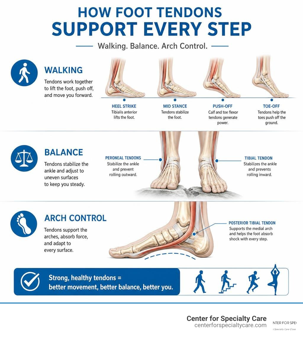
Foot tendons do three big things: they move the foot, stabilize the foot, and store or release energy. They are part of the larger kinetic chain that includes the ankle, knee, hip, pelvis, and spine.

How Tendons in the Foot Work During Walking and Running
During walking, the foot goes through phases:
- Heel strike: The foot contacts the ground. The anterior tibial tendon helps control how the foot lowers.
- Midstance: Body weight moves over the foot. The posterior tibial and peroneal tendons help stabilize the arch and ankle.
- Toe-off: The heel rises. The Achilles tendon, flexor hallucis longus, and toe flexors help generate push-off.
- Swing phase: The foot clears the ground. Extensor tendons lift the toes and the anterior tibial tendon lifts the foot.
Running increases the load. Tendons act like springs, storing energy as they stretch and releasing it as they recoil. That spring-like behavior improves efficiency, but it also means a sudden training jump can overload the system.
How Tendons Support the Medial, Lateral, and Transverse Arches
Your foot has multiple arches, not just one.
The posterior tibial tendon supports the medial arch. The peroneus longus helps stabilize the transverse arch across the midfoot. The peroneal tendons also support the lateral side of the foot. The plantar fascia, intrinsic foot muscles, and ligaments all share the load.
When one structure weakens, the others often compensate. Flat feet may overload the posterior tibial tendon. High arches may place more stress on the peroneal tendons. Tight calves may increase strain on the Achilles tendon and plantar fascia.
Research on the Plantar fascia and Achilles tendon relationship has shown that these structures are anatomically and functionally connected through tissues around the heel. That is one reason calf flexibility can affect heel and arch symptoms.
Why the Achilles Tendon and Plantar Fascia Are Often Linked
The plantar fascia is technically a ligament-like band, not a tendon, but it works closely with the foot tendon system. It runs along the bottom of the foot from the heel toward the toes and helps support the arch.
Tightness in the Achilles-calf complex can increase tension through the heel and plantar fascia. This is why heel pain treatment often includes both calf stretching and plantar fascia stretching. Night splints may help some patients by holding the Achilles tendon and plantar fascia in a lengthened position during sleep.
Structural research on Achilles-plantar fascia continuity has also described bridge-like collagen and bony relationships around the calcaneus. In plain English: the back of the heel and bottom of the heel talk to each other more than most people realize.
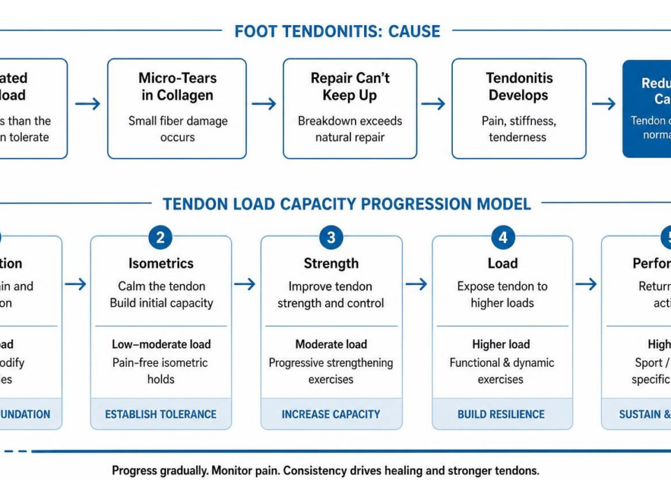
Common Foot Tendon Problems, Tendonitis Types, and Symptoms
Tendon pain may be described with several terms:
- Tendonitis: inflammation or irritation of a tendon.
- Tendinopathy: a broader term for painful tendon degeneration or overload.
- Tendinosis: chronic tendon wear without much active inflammation.
- Tenosynovitis: inflammation of the tendon sheath.
- Partial tear: some fibers are torn.
- Rupture: the tendon is completely torn.
For a broader overview, see our Foot tendonitis guide.
Achilles Tendonitis and Achilles Rupture
Achilles tendonitis often causes pain, swelling, warmth, thickening, and morning stiffness in the back of the heel or lower calf. Pain may be in the midportion of the tendon or where it inserts into the heel bone.
Achilles tendonitis affects an estimated 1% to 9% of elite and recreational athletes. It is common in runners, jumpers, and active adults who increase intensity too quickly.
A rupture is different. It may feel like a sudden pop, snap, or being kicked in the back of the ankle. Symptoms can include bruising, swelling, trouble walking, and inability to rise onto the toes. You can read more in our Achilles tendonitis insights.
Extensor Tendonitis on the Top of the Foot
Extensor tendonitis causes pain on the top of the foot, often worsened by tight shoes, tight laces, hill running, speed work, or repetitive toe lifting. The area may feel tender or swollen, and pain can increase when lifting the toes against resistance.
Changing footwear, loosening laces, reducing aggravating activity, and physical therapy often help. Our guide to Extensor tendonitis recovery covers this condition in more detail.
Posterior Tibial Tendonitis and Adult Acquired Flatfoot
Posterior tibial tendonitis usually causes pain and swelling along the inside of the ankle or arch. Tenderness near the navicular bone is common. Patients may notice increasing flattening of the arch, overpronation, or the “too-many-toes” sign, where more toes are visible from behind because the forefoot drifts outward.
A single-leg heel raise is often difficult or painful. If the tendon continues to weaken, adult acquired flatfoot can progress from flexible arch collapse to a more rigid deformity.
Peroneal Tendonitis, Instability, and Split Tears
Peroneal tendon problems cause pain behind or below the outside ankle bone. Symptoms may include swelling, clicking, weakness with eversion, or a sense that the ankle is unstable.
High arches, repeated ankle sprains, uneven surfaces, and sudden side-to-side sports can contribute. The peroneus brevis is especially known for split tears. Sometimes the tendons sublux, meaning they slip out of their normal groove behind the ankle.
Flexor and Plantar-Side Tendon Disorders
Flexor hallucis longus tendonitis can cause pain behind the ankle, under the arch, or near the big toe. It is sometimes seen in dancers and athletes who repeatedly push off through the big toe.
Flexor digitorum longus problems may cause pain along the bottom of the foot or with toe gripping. Plantar tendon disorders can be difficult to distinguish from nerve irritation, plantar plate injuries, sesamoid pain, and plantar fasciitis. Imaging can be especially useful here. A radiology review of MR and ultrasound evaluation of plantar foot tendons explains how detailed imaging helps identify tendinosis, tenosynovitis, partial tears, complete tears, entrapment, and instability.
How Foot Tendon Injuries Are Diagnosed and Treated
Accurate diagnosis starts with listening. We want to know when symptoms started, what changed in activity, what shoes you wear, whether there was a pop or injury, and what makes the pain better or worse.
Physical Exam Tests for Tendons in the Foot
A foot tendon exam may include:
- Palpation to map tenderness and swelling.
- Range-of-motion testing at the ankle, foot, and toes.
- Resisted strength testing for dorsiflexion, plantar flexion, inversion, eversion, and toe motion.
- Single-leg heel raise to assess Achilles and posterior tibial function.
- Thompson test for suspected Achilles rupture.
- Gait analysis to see how the foot loads during walking.
- Arch assessment while sitting, standing, and rising onto the toes.
- Ankle stability testing, especially for peroneal tendon symptoms.
- Neurovascular check to make sure circulation and nerve function are intact.
The location of pain matters. Back of heel, inside ankle, outside ankle, top of foot, and bottom of forefoot often point toward different tendons.
Imaging for Foot Tendon Pain
Imaging depends on the suspected problem.
X-rays do not show tendons directly, but they can show bone spurs, arthritis, fractures, alignment problems, heel spurs, and flatfoot changes.
Ultrasound can show tendon thickening, fluid around a tendon sheath, partial tears, and movement problems. Dynamic ultrasound is helpful because the tendon can be observed while the foot moves.
MRI provides detailed images of tendons, bones, joints, ligaments, and soft tissue. It is commonly used when a tear, rupture, chronic tendinopathy, or complex diagnosis is suspected.
CT is less common for tendon diagnosis but may help when bone alignment, fractures, or surgical planning are part of the concern.
Non-Surgical Treatment Options
Most foot tendon problems begin with conservative care. Depending on the diagnosis, treatment may include:
- Rest or activity modification.
- Ice for pain and swelling.
- Compression and elevation.
- Anti-inflammatory medication when appropriate.
- Supportive shoes.
- Custom or over-the-counter orthotics.
- A walking boot for more painful cases.
- Bracing for ankle instability or tendon protection.
- Physical therapy.
- Calf stretching and plantar fascia stretching.
- Eccentric strengthening, especially for Achilles tendinopathy.
- Balance training and gait retraining.
- Gradual return to sport or work activity.
Physical therapy is often the turning point. Tendons generally dislike sudden change, but they respond well to progressive loading when guided correctly. Our article on Physical therapy and recovery information discusses healthy-foot strategies and treatment options.
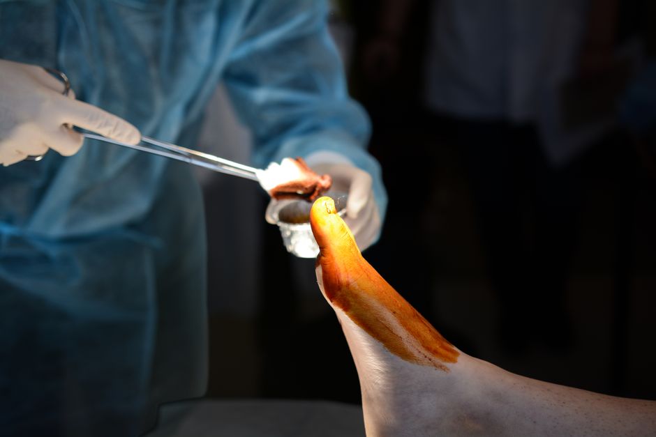
Surgical Treatment Options for Severe Tendon Disorders
Surgery is not the first step for most patients, but it may be needed for complete ruptures, severe tears, progressive deformity, or symptoms that do not improve with appropriate non-surgical care.
Surgical options may include:
- Tendon repair for torn tendon fibers.
- Debridement to remove damaged tendon tissue.
- Tenosynovectomy to remove inflamed tendon sheath tissue.
- Tendon transfer when one tendon must help replace another.
- Achilles rupture repair.
- Gastrocnemius recession for severe calf tightness contributing to tendon overload.
- Flatfoot reconstruction for advanced posterior tibial tendon dysfunction.
- Peroneal tendon stabilization or groove deepening for recurrent subluxation.
- Repair of peroneal split tears.
Recovery usually involves a boot or cast, a period of protected weight-bearing, and progressive rehabilitation. Complex tendon surgery may take 6 to 12 months for full recovery. Our guide to Podiatric surgery expectations explains what patients can generally expect before and after foot procedures.
Prevention, Long-Term Prognosis, and When to See a Specialist
The best tendon injury is the one you never have to treat. Prevention is not glamorous, but neither is limping across a parking lot because your Achilles is angry.
Prevention Strategies for Active Adults and Athletes
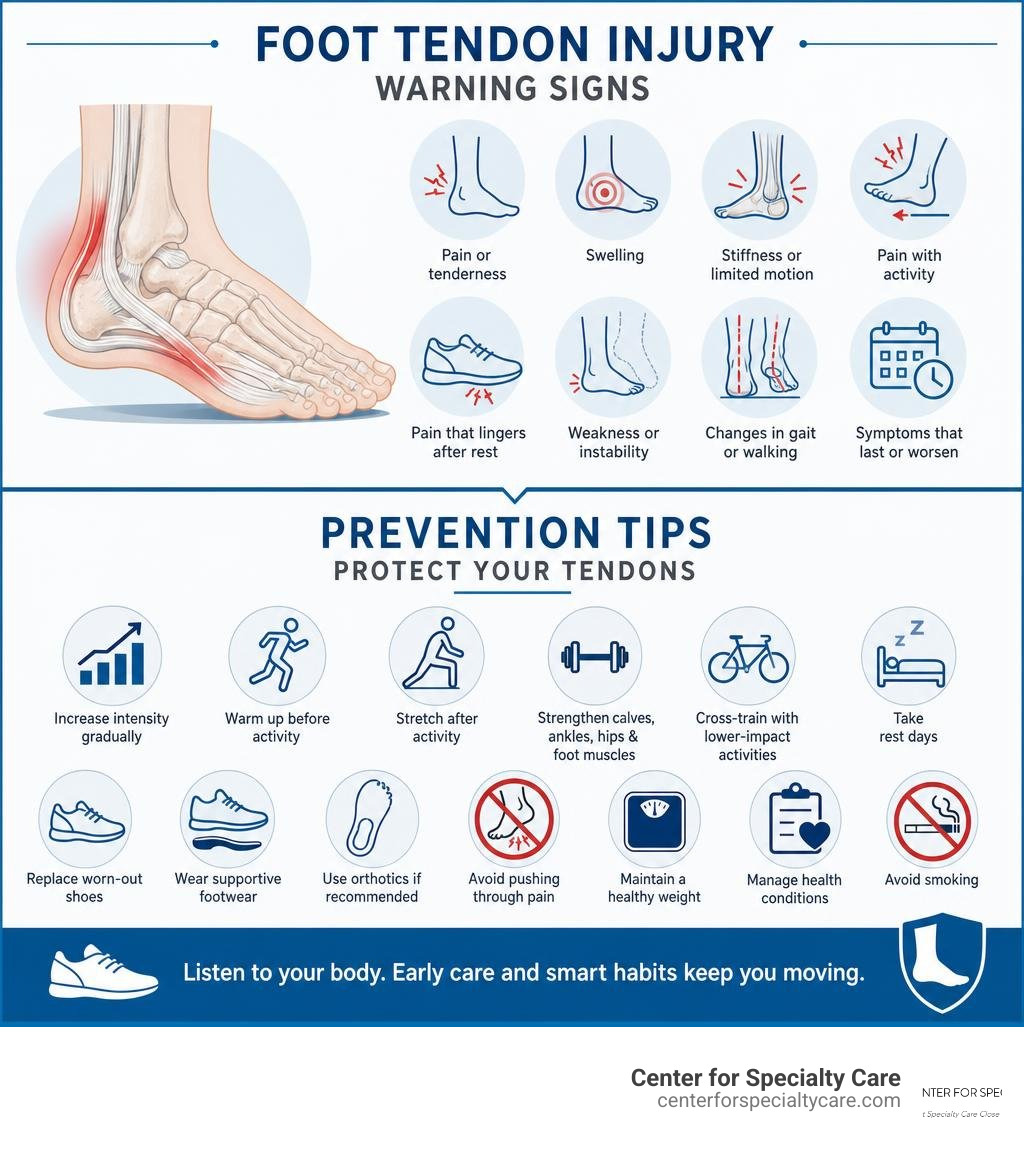
To reduce your risk of foot tendon problems:
- Increase mileage, speed, hills, or sports intensity gradually.
- Warm up before activity.
- Stretch calves and feet after activity.
- Strengthen calves, ankles, hips, and intrinsic foot muscles.
- Cross-train with lower-impact activity.
- Take rest days.
- Replace worn-out shoes.
- Use supportive footwear for your foot type.
- Use orthotics if recommended.
- Avoid pushing through sharp or worsening tendon pain.
- Maintain a healthy weight.
- Manage diabetes, arthritis, gout, thyroid disease, and other conditions that can affect tendon health.
- Avoid smoking, which can impair tissue healing.
Rehabilitation Exercises That Support Recovery
Rehab should be matched to the diagnosis, but common exercises include:
- Calf raises for Achilles and ankle strength.
- Eccentric heel drops for Achilles tendinopathy when appropriate.
- Towel curls for toe flexor and intrinsic foot strength.
- Resisted inversion for posterior tibial strengthening.
- Resisted eversion for peroneal strengthening.
- Toe yoga to improve big-toe control and arch coordination.
- Single-leg balance drills.
- Plantar fascia stretching.
- Wall calf stretches with knee straight and knee bent.
- Gradual walking or running progression.
A key reminder: tendon rehab should create controlled challenge, not a pain explosion. Mild soreness can happen, but sharp pain, swelling, or next-day worsening means the load may be too high.
Long-Term Outlook After Foot Tendon Injuries
Many people recover fully from foot tendonitis with early care, proper footwear, activity changes, and rehabilitation. Mild to moderate tendonitis may improve over weeks to months, while chronic tendinopathy takes longer because tendon remodeling is slow.
The outlook depends on:
- Which tendon is involved.
- Whether there is a tear or rupture.
- How long symptoms have been present.
- Foot structure, such as flat feet or high arches.
- Training load and work demands.
- Medical conditions that affect healing.
- Consistency with rehab.
Reinjury is possible, especially if someone returns to high activity too quickly. Long-term maintenance exercises and smart footwear often make the difference between “I got better” and “Here we go again.”
When Foot Tendon Pain Needs Medical Care
Do not ignore these warning signs:
- A snap, pop, or sudden tearing sensation.
- Inability to walk normally.
- Severe swelling or bruising.
- Inability to rise onto the toes.
- New foot deformity or worsening arch collapse.
- Pain that persists despite rest and home care.
- Numbness, tingling, or color change.
- Redness, warmth, fever, or concern for infection.
- Recurrent ankle instability or tendon clicking.
- Pain that keeps returning when you resume activity.
If you suspect an Achilles rupture or another major tendon injury, prompt evaluation is important. Waiting can make treatment more complicated.
Conclusion
The tendons in the foot are small compared with many other body structures, but their job is enormous. They help you walk, balance, climb, run, pivot, and stand through thousands of steps every day.
At Center for Specialty Care, we provide personalized evaluation and treatment for foot and ankle pain, including both non-surgical and surgical options when appropriate. Our team serves patients in Fairmont, Minnesota, Estherville, Iowa, Buffalo Center, Iowa, St. James, Minnesota, and surrounding Minnesota and Iowa communities.
If foot tendon pain is limiting your work, workouts, or daily life, we are here to help with quick appointment availability, individualized care, and a commitment to patient satisfaction.
To take the next step, Schedule care for foot pain and tendon problems.