
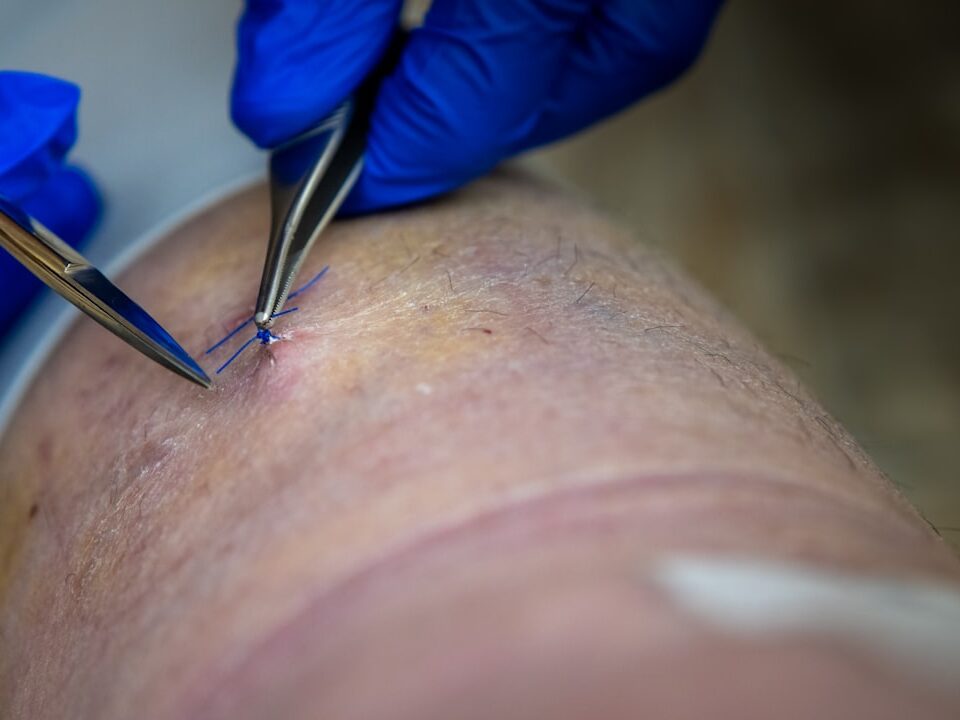
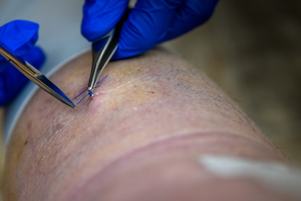
Beyond the Tear: Effective Treatments for Rotator Cuff Injuries
September 26, 2025
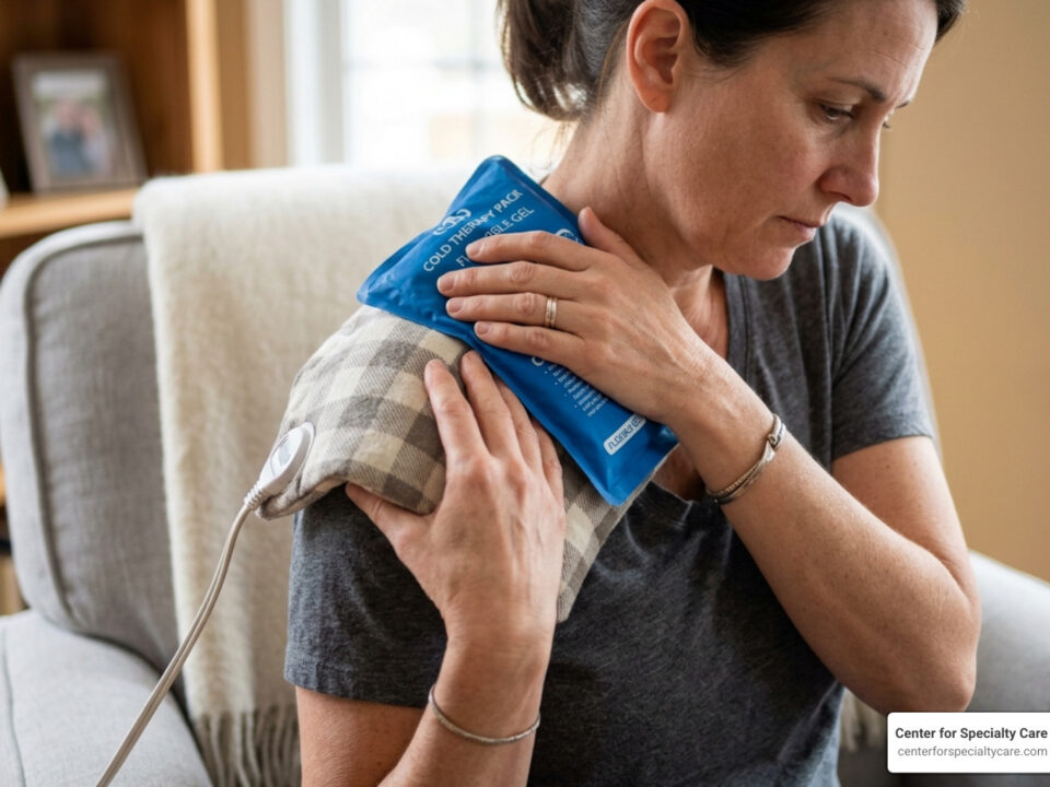
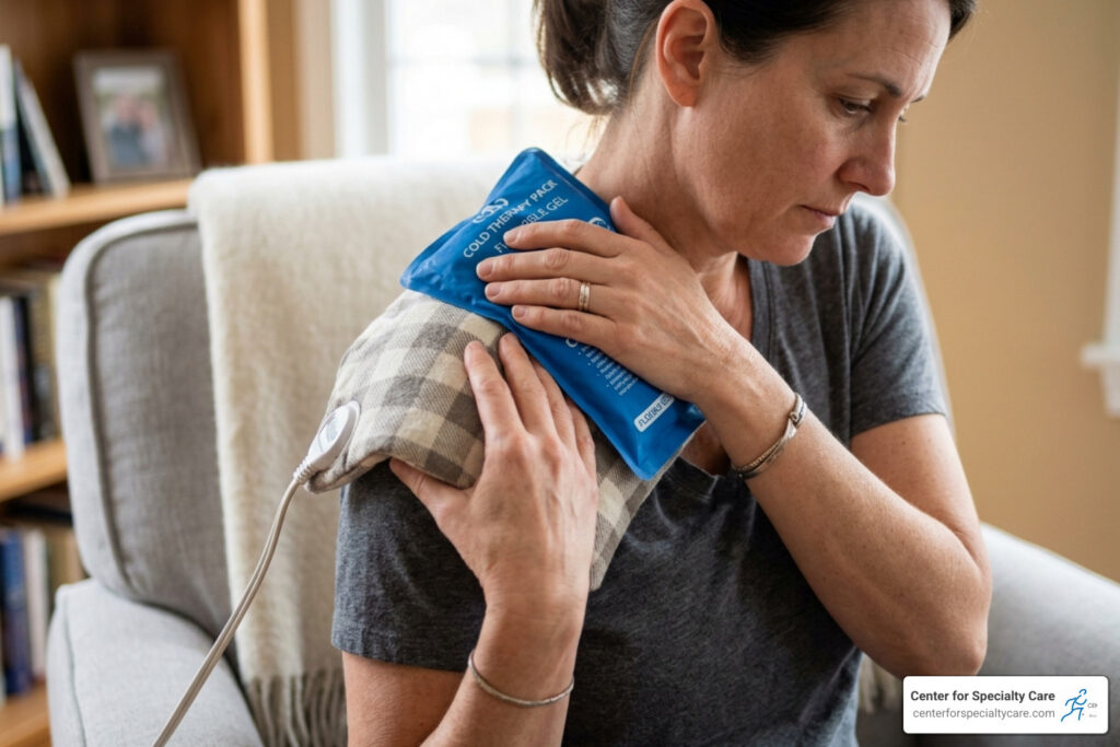
How to Get Rid of Shoulder Blade Muscle Knots
September 30, 2025
Diagram of a shoulder: Top 1 Expert Guide
The Shoulder’s Complex Engineering Made Simple
A diagram of a shoulder reveals one of the body’s most remarkable joints. Understanding its structure helps you care for your shoulders and spot problems early.
Key Components of a Shoulder Diagram:
- Three main bones: Humerus (upper arm), scapula (shoulder blade), and clavicle (collarbone)
- Four joints: Glenohumeral (main ball-and-socket), acromioclavicular (AC), sternoclavicular (SC), and scapulothoracic
- Rotator cuff muscles: Four muscles (supraspinatus, infraspinatus, teres minor, subscapularis) that stabilize the joint
- Supporting structures: Ligaments, labrum, bursae, cartilage, nerves, and blood vessels
This ball-and-socket joint gives you the greatest range of motion in the body, but that mobility trades off with stability. The humeral head is much larger than the shallow socket—think a golf ball on a tee—so muscles and soft tissues must keep it centered.
As Dr. Corey Welchlin, a board‑certified orthopedic surgeon, I’ve seen patient education make a real difference: understanding how a diagram of a shoulder relates to your symptoms is the first step toward better outcomes.

Diagram of a shoulder definitions:
- partially dislocated shoulder symptoms
- shoulder feels out of place but not dislocated
- what does a dislocated shoulder look like
A Detailed Diagram of a Shoulder: The Skeletal Framework
Your shoulder starts with three key bones: the humerus (upper arm), scapula (shoulder blade), and clavicle (collarbone). Together they form the shoulder girdle, the flexible connection between your arm and torso.

Each bone has a distinct role: the humerus provides the “ball,” the scapula forms the shallow “socket” and broad muscle attachment surfaces, and the clavicle acts like a strut that positions the shoulder for motion and strength. Their precise articulation enables everyday activities from reaching overhead to throwing.
The scapula itself is a complex triangular bone with multiple important landmarks. The acromion process forms the roof of the shoulder, the coracoid process serves as an attachment point for several muscles and ligaments, and the glenoid cavity creates the socket for the humeral head. Understanding these bony landmarks helps explain why certain movements or positions may cause discomfort.
For comprehensive joint care, explore our orthopedics services.
The Glenohumeral Joint: The Main Ball-and-Socket
The glenohumeral joint—the classic ball-and-socket—is highly mobile because the glenoid socket covers only a fraction of the humeral head. Both articular surfaces are lined with hyaline cartilage and surrounded by a capsule filled with synovial fluid for low-friction motion. This remarkable design delivers motion but demands strong soft-tissue support.
The joint capsule itself is twice the surface area of the humeral head, allowing for extensive movement. This redundancy in the capsule creates recesses that unfold during different arm positions, enabling the shoulder’s remarkable flexibility. The synovial membrane within produces fluid that nourishes the cartilage and reduces friction, maintaining smooth motion throughout decades of use. Learn more about the anatomy of the shoulder joint.
The Acromioclavicular (AC) and Other Key Joints
Your shoulder complex includes three additional joints that coordinate motion:
- Acromioclavicular (AC) joint: links the acromion to the clavicle and stabilizes overhead motions. This small joint bears significant stress during lifting and carrying activities.
- Sternoclavicular (SC) joint: the only bony connection of the arm to the torso. Despite its importance, this joint rarely causes problems due to its strong ligamentous support.
- Scapulothoracic articulation: the shoulder blade glides over the rib cage, enabling full arm elevation. This “pseudo-joint” contributes about one-third of total shoulder motion.
When any of these joints are limited, the others compensate, often provoking symptoms. The synchronized movement of all four joints creates what experts call scapulohumeral rhythm—a precise coordination that allows smooth, powerful arm movement.
The Soft Tissues That Support and Move the Shoulder
Bones are the frame; soft tissues power and protect the joint. Muscles move the arm, tendons connect muscles to bone, ligaments limit excessive motion, and cartilage cushions contact surfaces.

Visualizing the Rotator Cuff on a Diagram of a Shoulder
The rotator cuff—”SITS”: Supraspinatus, Infraspinatus, Teres minor, Subscapularis—wraps the humeral head like a cuff. Beyond moving the arm (initiation of abduction, external and internal rotation), its primary job is dynamic stabilization: keeping the ball centered in the shallow socket during every motion. If these muscles weaken or tear, motion becomes painful and less controlled. Find expert options in our rotator cuff surgeon guide.
The Stabilizing Role of Ligaments, Labrum, and Bursae
Ligaments (especially the glenohumeral and coracohumeral) reinforce the capsule and act like seat belts to limit excessive translation. The glenoid labrum deepens the socket and anchors key ligaments, improving stability. The joint capsule is loose enough to allow motion yet strong enough to contain the joint. Bursae, including the subacromial bursa, reduce friction where tendons pass under bony roofs.

Nerves and Blood Supply
The brachial plexus supplies shoulder function. The axillary nerve powers the deltoid and provides sensation over the lateral shoulder—often checked after dislocations. The suprascapular nerve powers supraspinatus and infraspinatus. Arterial supply, including the circumflex humeral arteries, nourishes bone and soft tissue for performance and healing.
How the Shoulder Moves: A Symphony of Motion
The shoulder moves through multiple planes: flexion (forward/up to 180 degrees), extension (backward to 60 degrees), abduction (out to the side to 180 degrees), adduction (back to the body, crossing midline up to 50 degrees), internal rotation (up to 90 degrees), external rotation (up to 90 degrees), and combined circumduction. These movements rarely occur in isolation—most daily activities involve complex combinations.
Abduction starts with the supraspinatus initiating the first 15-30 degrees, then the deltoid takes over as the primary mover. Full elevation requires coordinated scapular rotation by the trapezius and serratus anterior. The rotator cuff stabilizes throughout while larger muscles (deltoid, pectoralis major, latissimus dorsi) produce power. This intricate timing, called force coupling, ensures smooth motion without impingement.
During overhead reaching, the scapula must rotate upward, tilt posteriorly, and externally rotate—all while the clavicle lifts and rotates backward. If stiffness or pain disrupts this timing, compensatory patterns develop that can lead to secondary problems. Targeted therapy can restore proper movement patterns and prevent long-term issues. See our approach to shoulder treatment with physical therapy.
The Trade-Off: Why High Mobility Can Lead to Instability
A shallow glenoid and a relatively loose joint capsule permit exceptional mobility but reduce inherent stability. The glenoid cavity covers only about 25-30% of the humeral head at any given position. Soft tissues—especially the rotator cuff—must constantly work to keep the humeral head centered.
This dynamic stabilization system relies on proprioception (position sense) and coordinated muscle firing. When these muscles fatigue or are injured, the shoulder becomes vulnerable, and dislocation risk rises. The most common position for dislocation is with the arm abducted and externally rotated—the “high five” position—where the anterior capsule is most stressed. Athletes in overhead sports face particular challenges maintaining this delicate balance between mobility and stability. More on anatomy and common injuries.
When Things Go Wrong: Common Shoulder Conditions
With so many moving parts, shoulder issues are common—from acute injuries to age‑related wear. The upside: most problems respond well to early, appropriate care.

Understanding the Most Common Shoulder Injuries
Rotator cuff tears: From overuse or trauma. Symptoms include deep ache, weakness with lifting, and night pain. Tears may be partial or full‑thickness.
Shoulder impingement: Rotator cuff tendons are pinched during elevation, often causing a “painful arc” around shoulder height.
Shoulder dislocations: The humeral head displaces from the socket—usually anteriorly—creating intense pain and future instability risk.
Bursitis: Inflamed bursae cause pain with motion or lying on the shoulder due to increased friction.
Arthritis: Cartilage wear leads to stiffness, pain, and possible grinding (crepitus).
Labral tears (including SLAP): Damage to the labrum causes deep pain, clicking, and a sense of giving way—common in throwers and lifters.
If persistent pain limits your activities, we can help identify the source and guide treatment. Learn more about shoulder joint pain.
Why Is My Shoulder Clicking or Popping?
Occasional painless pops can be benign (gas bubble release). Painful or recurrent clicking often signals a problem:
- Labral tears can cause catching as the head moves against a torn rim.
- Inflamed bursae or tendons create grinding or creaking when tissues no longer glide smoothly.
- Arthritic cartilage wear causes rough surfaces and crepitus.
- Loose bodies can intermittently catch and lock the joint.
- Ligament laxity can allow excess motion and popping.
If noises are frequent or painful, an evaluation can prevent worsening. Read more about why your shoulder is popping.
Frequently Asked Questions About Shoulder Anatomy
Patients viewing a diagram of a shoulder often ask similar questions. Clear answers make anatomy—and recovery—easier to understand.
What are the four main muscles of the rotator cuff?
The cuff includes the supraspinatus, infraspinatus, teres minor, and subscapularis. They form a stabilizing sleeve around the humeral head and enable arm rotation and the start of abduction.
What is the labrum and why is it important?
The labrum is a fibrocartilage rim that deepens the shallow glenoid, improves suction stability, and anchors key ligaments—essential for keeping the humeral head centered, especially during overhead activity.
What is the difference between a tendon and a ligament in the shoulder?
Tendons connect muscle to bone (e.g., rotator cuff tendons) to create movement. Ligaments connect bone to bone (e.g., glenohumeral ligaments) to limit excessive motion and provide static stability.
Your Guide to Comprehensive Shoulder Care
Every reach, lift, and throw relies on a finely tuned system: the glenohumeral joint for motion, the rotator cuff for stability, the labrum and ligaments for support, and smooth soft‑tissue gliding for comfort. That mobility also means the shoulder is prone to overuse and injury—knowledge and early care matter.
At Center for Specialty Care, we focus on 100% patient satisfaction with personalized, conservative-first treatment and quick appointment availability across our Minnesota and Iowa locations. Whether you need education, physical therapy, injections, arthroscopy, or joint replacement, we tailor care to your goals so you can return to what you love.
